IFRM 2025 Workshop
Intro
The UIC Institute for Functional and Regenerative Materials (IFRM) is thrilled to announce a dynamic workshop focused on the cutting-edge fields of functional imaging and functional biomaterials. This event is designed to bring together scientists, researchers, and professionals with a shared interest in these innovative areas to foster learning, networking, and collaboration.
Workshop Details
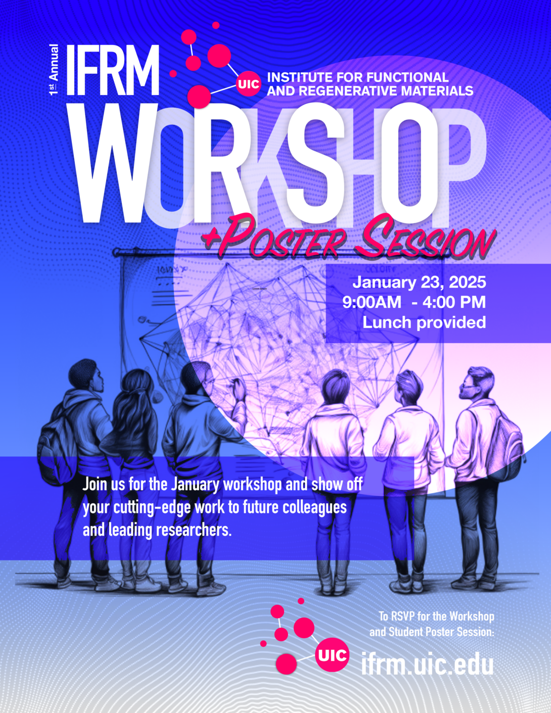
January 23, 2025 // 9:00 AM – 4:00 PM
UIC Herman Auditorium (Room 1017)
Molecular Biology Research Building
About the Workshop
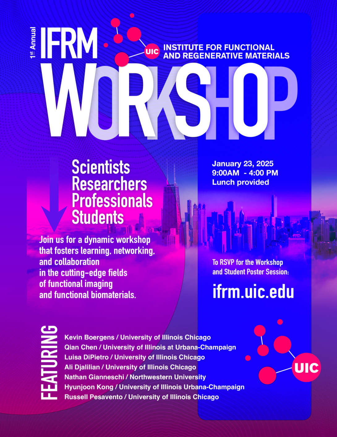
Over the past 50 years, new materials have permeated virtually every aspect of society, revolutionizing our communication, transportation, agricultural and healthcare sectors. This workshop focuses on two aspects of materials science. Functional imaging involves developing new advanced microscopy techniques to characterize the structure of adaptive, smart, and biological materials. In contrast, functional biomaterials involves the development of novel materials that will interface with biological systems to improve tissue and organ function. The workshop will provide a forum for interaction between medical, engineering, and physical science research groups.
Join the IFRM for expert speakers, networking opportunities, and to explore new collaborations. Hear from leading scientists who are shaping the future of functional imaging and functional biomaterials, gaining insights into the latest research, technologies, and breakthroughs. Connect with peers and speakers in a stimulating environment designed to spark new ideas and partnerships. Discover future research and development opportunities through networking sessions and collaborative discussions.
Scientists, researchers, and graduate students eager to expand their knowledge and connect with a broader network of researchers in the fields of functional imaging and biomaterials are welcome join us at no cost — and enjoy a free lunch!
Workshop Agenda
| 9:00 AM | Welcome |
| 9:05 AM | Nathan Gianneschi (Northwestern University) |
| 9:45 AM | Qian Chen (University of Illinois Urbana-Champaign) |
| 10:25 AM | Kevin Boergens (University of Illinois Chicago) |
| 10:50 AM | Russell Pesavento (University of Illinois Chicago) |
| 11:10 AM | Poster Session |
| 12:00 PM | Lunch |
| 1:00 PM | Hyun Joon Kong (University of Illinois Urbana-Champaign) |
| 1:45 PM | Georgia Papavasiliou (Illinois Institute of Technology) |
| 2:25 PM | Luisa DiPietro (University of Illinois Chicago) |
| 2:50 PM | Ali Djalilian (University of Illinois Chicago) |
| 3:15 PM | PI Roundatble with IFRM |
| 3:45 PM | Conclusion |
Invited Speakers and Abstracts
Photoemission electron microscopy for high-throughput biological imaging
Abstract
High-throughput biological imaging is critical for advancing our understanding of complex systems, from the brain’s intricate networks to diverse biological and material structures. We are developing a Photoemission Electron Microscope (PEEM) that has the potential to dramatically increase imaging throughput while reducing costs. PEEM combines the reliability of Scanning Electron Microscopy (SEM) with the speed of Transmission Electron Microscopy (TEM), enabling rapid, high-resolution imaging. Our PEEM system incorporates UV laser excitation and wide-field electron detectors, making imaging rates on the scale of gigahertz plausible. These capabilities have already been demonstrated to visualize synapses, an important milestone for connectomics. Beyond neural applications, this technology shows promise for studying nanoscale biological processes such as biomineralization, where imaging of complex interfaces is essential. To realize the potential of PEEM, we are optimizing the entire imaging pipeline. This includes advancing ultra-thin section collection with automated techniques like ATUM and Mag-C, improving Z-resolution using Gas Cluster Ion Beam milling, and developing fast mechanical stages to match our imaging rates. Automation plays a key role, with integrated auto-focus, auto-contrast, and quality assurance algorithms ensuring consistent data acquisition across thousands of sections. This talk will explore the transformative implications of PEEM microscopy, from accelerating discoveries in neuroscience to opening new avenues for nanoscale imaging in biology. By enhancing both the speed and accessibility of imaging, this approach could push high-throughput biological imaging forward.
About Dr. Boergens / Profile
University of Illinois Chicago
Kevin Boergens is a Research Assistant Professor in the Departments of at the University of Illinois Chicago. Kevin came to UIC from Austin, Texas where he worked in industry at Paradromics Inc. to build high data-rate brain computer interfaces (BCI). His research interests at UIC include using 3D electron microscopy to map brain circuits. His newest efforts focus on developing photoemission electron microscopy as a tool for neuroscience. Kevin received his doctorate from the Ludwig Maximilian University of Munich.
Electron videography and Its automation for soft materials
Abstract
My group studies the spatiotemporal heterogeneity of materials and addresses the associated science and technological questions of how to image it, quantify it, understand it, and engineer it for new properties, from the finest atomic and molecular scale to the particle assembly and composite scale. Specifically, we adapt a suite of electron videography methods (e.g., liquid-phase TEM, electron tomography, 4D-STEM) and machine-learning based data-mining to synthetic soft, biological, and energy related systems. In today’s talk, I will focus on the synthesis and phase behaviors of colloidal nanoparticles. We image the crystallization pathways in solution, examining effects such as discrete molecular interaction and multi-scale coupling, which complicate the potential energy surface. By combining single particle tracking and simulations, we discover interesting chemical phenomena, such as non-classical nucleation, size-dependent crystal growth habits, and moiré patterning, enabling advanced crystal engineering. We will emphasize our efforts in automated data acquisition and analysis, often aided with machine learning, to allow us to perform statistically-significant analysis, to understand the presence and importance of spatiotemporal heterogeneity in morphology, composition, structure, and function.
Qian Chen / Profile
University of Illinois at Urbana-Champaign
Qian Chen is currently a Professor and Racheff Scholar in the Materials Science and Engineering Department at University of Illinois at Urbana-Champaign (UIUC). She obtained her PhD from the same department and completed her postdoc at UC Berkeley as a Miller Fellow. She joined the faculty of UIUC in 2015 and since then has received awards for the research in her group including the Victor LaMer Award in ACS, made the Forbes 30 under 30 Science List, the Air Force Office of Scientific Research YIP Award, a National Science Foundation CAREER Award, was named a Sloan Research Fellow in Chemistry, the Unilever Award in ACS, the UIUC Dean’s Award for Excellence in Research (twice), and the Hanwha-TotalEnergies IUPAC Young Scientist Award. She and her research group focus on the broad scheme of imaging, understanding and engineering soft materials at the nanoscale, including systems such as colloidal self-assembly, protein transformation, advanced battery devices, metamaterials, and energy-efficient separation strategies.
Wound complexity: Challenges in the development of effective therapeutic materials
Abstract
The ability to adequately repair injured tissue is essential to human health, and the clinical problem of nonhealing wounds is a serious and growing problem. Despite tremendous advances in our understanding of the biological complexity of the repair process, the translation of this knowledge into effective treatments for wounds has been slow and difficult. This lecture will discuss the obstacles that must be overcome to develop new and beneficial materials for wound therapeutics.
About Dr. DiPietro / Profile
University of Illinois Chicago
Dr. Luisa A. DiPietro is Professor of Periodontics and Director of the UIC Center for Wound Healing and Tissue Regeneration. Dr. DiPietro received both her DDS and PhD in immunology from the University of Illinois Chicago, and completed a residency in hospital dentistry at Michael Reese Hospital in Chicago. Dr. DiPietro’s research focuses on wound healing, and she has been the recipient of more than $19 million in extramural research grants and contracts. Her work has been cited more than 31,000 times. Her honors and awards include the Lifetime Achievement Award from the Wound Healing Society, the Mentor of the Year Award from the American Association for Dental Research Student Research Group, and the University of Illinois – University Scholar Award. Dr. DiPietro is an elected Fellow of the American Association for the Advancement of Science and a member of the National Advisory Dental and Craniofacial Research Council for the National Institutes of Health.
Biomaterial and cell based approaches for corneal repair and regeneration
Abstract
The cornea is an optically clear and avascular tissue that is central for vision. In addition to specialized cells, it is largely composed of a highly organized extracellular matrix. Our studies have focused on cell and biomaterial based approached to promote regenerative healing in the cornea. This has application for severe corneal diseases where corneal wound healing is impaired.
About Dr. Djalilian / Profile
University of Illinois Chicago
Ali Djalilian, MD is the Searls-Schenk Professor of Ophthalmology at University of Illinois Chicago. He is a cornea specialist and a clinician-scientist who balances his time between seeing patients and leading a laboratory and translational research program funded by NIH and Department of Defense focused on developing novel regenerative therapies for the cornea.
Liquid phase TEM for structure elucidation and property prediction of dynamic biomaterials
Abstract
Organic, soft materials with nanoscale structures inherent to their solvated state, such as emulsions, hydrogels, biomaterials and thermally responsive materials, provide fertile ground for investigation via direct imaging using liquid phase transmission electron microscopy (LPTEM). With key advances having been made in the past decade, including improved sample preparation protocols, image capture technologies, and image analysis, LPTEM has gained in utility to the point where we are able to investigate materials that are otherwise difficult or impossible to directly image by other methods with nanoscale resolution. This workflow consists of (1) modelling electron beam-solvent interactions, (2) studying electron beam-sample interactions via LCTEM coupled with post-mortem analysis, (3) construction of “damage plots” displaying sample integrity under varied imaging and sample conditions, (4) optimized LCTEM imaging, (5) image processing, and (6) correlative analysis via X-ray or light scattering. Herein, we describe this workflow for the examination of biomolecular hydrogels including their dynamics and assembly in the solvated state. The insight gained has provided an exciting window into these materials that eludes cryogenic TEM, which is necessarily disruptive of these structures. We present our efforts in this space, and in the correlative methods used to verify, or support the structural information found. This includes the use of LPTEM to define gel parameters including persistence length and mesh sizes, which has led to the prediction of properties in thermally responsive gels. We will also give some perspective on how we leverage these capabilities for other materials with relevance to biomedical materials we are developing in regenerative medicine, cancer, dermatology and neurodegenerative disease.
About Dr. Gianneschi / Profile
Northwestern University
Nathan C. Gianneschi received his B.Sc(Hons) at the University of Adelaide, Australia in 1999 under Louis Rendina. In 2005 he completed his Ph.D at Northwestern University, with Chad Mirkin. Following a Dow Chemical postdoctoral fellowship at The Scripps Research Institute with Reza Ghadiri, in 2008 he began his independent career at the University of California, San Diego where, until June 2017, he was Teddy Traylor Scholar and Professor of Chemistry & Biochemistry, NanoEngineering and Materials Science & Engineering. In July of 2017, Gianneschi moved his research group to Northwestern University where he is currently Jacob & Rosaline Cohn Professor of Chemistry, Materials Science & Engineering, and Biomedical Engineering. Gianneschi takes an interdisciplinary approach to nanomaterials research with a focus on multifunctional materials with interests that include biomedical applications, programmed interactions with biomolecules and cells, and basic research into nanoscale materials design, synthesis and characterization, including via new electron microscopy methods. Key areas of focus include the development of mimetic biological materials through chemical and nanoscale synthesis, including the development of protein mimetics and mimetics of melanin pigments. Gianneschi is co-Founder of multiple start-up companies pursing clinical translation of these technologies, including for the treatment of cancer. For this work he has been awarded the NIH Director’s New Innovator Award, the NIH Director’s Transformative Research Award and the White House’s highest honor for young scientists and engineers with a Presidential Early Career Award for Scientists and Engineers (AFOSR). Prof. Gianneschi was awarded a Dreyfus Foundation Fellowship, is a Kavli Fellow of the National Academy of Sciences, a Fellow of the Royal Society of Chemistry, and is an Alfred P. Sloan Foundation Fellow.
Reviving diatoms: A new strategy for biofilm elimination
Abstract
Biofilms are bacterial communities encased in extracellular polymeric substances (EPS), which serve as a protective barrier. These biofilms can infect biological tissues, foul biomedical devices, and even degrade infrastructure, posing significant threats to human health and sustainability. Compared to planktonic bacteria, biofilms exhibit high resistance to antibiotics due to the shielding effects of EPS. To address this challenge, we developed a novel self-propelling particle system designed to invade, damage, and ultimately remove biofilms. This system works by generating oxygen bubbles that burst with force, disrupting the biofilm structure. The particles are assembled by doping naturally derived cylindrical diatoms with catalysts capable of decomposing hydrogen peroxide into oxygen. This talk will explore the chemistry behind the fabrication of these diatom-based microbubbles and explain the mechanism by which they disrupt 3D biofilms, using cell-matrix analysis and optical coherence tomography. Additionally, we will demonstrate the system’s cleaning efficacy on biofilms contaminating complex surfaces such as surgical tools, dental implants, and biological tissues. Overall, this study represents a significant advance in biofilm management, offering a promising approach for combating biofilm-related infections and contamination.
About Dr. Kong / Profile
University of Illinois Urbana-Champaign
Hyunjoon Kong is a Robert W. Schafer professor in the Department of Chemical and Biomolecular Engineering, Carle Illinois College of Medicine, and Pathobiology at the University of Illinois at Urbana-Champaign (UIUC). He received his engineering education from the University of Michigan at Ann Arbor (Ph. D.) and performed post-doctoral research at the University of Michigan and Harvard University. He joined the University of Illinois in 2007. He received the NSF Career Award, the Center for Advanced Study Fellowship, the UIUC Engineering Dean’s Award for Research Excellence, the Centennial Scholar, and the Promotion Award. He was elected an American Institute of Medical and Biological Engineering (AIMBE) Fellow. To date, he has published more than 200 papers in various peer-reviewed journals. He is currently leading a multi-cellular engineered living systems (MCELS) theme in the Institute for Genomic Biology of UIUC. He also serves as an editorial board member of Biomaterials and Biofabrication journals and an associate editor of Biomaterials Research.
Hydrogel biomaterials: Advancing regenerative medicine and drug delivery
Abstract
Throughout the last decade our lab has focused on designing pro-angiogenic hydrogel materials to promote neovascularization of engineered tissues and hydrogel nanoparticles for therapeutic drug delivery. In this regard we have desiged vascular spheroid-laden poly(ethylene) glycol (PEG) hydrogel scaffolds to evaluate the impact of extracellular matrix cues on neovascularization in 3D culture and following subcutaneous implantation in vivo. We have utilized free-radical photopolymerization chemistries to create PEG diacrylate hydrogel scaffolds with highly tunable variations in modulus, proteolytic degradation rate and integrin binding cell adhesion peptide ligand concentration. Our studies have shown that synergistic enhancements in rates of proteolytic scaffold degradation and cell adhesive ligand concentration enhance vascular sprouting responses in 3D culture over a broad range of modulus of soft tissues. We have also recently developed a process for production of shelf-stable, fully biocompatible hydrogel-based NP emulsions (BCNE), suitable for en masse use to deliver hydrophilic therapeutics of varying size and conformation. This is achieved by optimizing hydrogel nanoparticle network mesh dimensions relative to the size and conformation of the encapsulated therapeutic to control its therapeutic release. In this presentation I will highlight some of our recent projects that utilize our BCNE platform (1) for intra-articular injection and (2) oral administration of therapeutics for targeted delivery to joint and gut tissues to attenuate angiogenesis and fibrosis as well as epithelial barrier dysfunction associated with rheumatoid arthritis, and (3) as topical pro-angiogenic ointments for healing of diabetic wounds. In vitro data involving repurposed FDA therapeutics to attenuate fibrosis and angiogenesis in 3D spheroid culture models as well as in vivodata on diabetic wound closure rate following topical application of pro-angiogenic peptide BCNE ointments will be presented.
About Dr. Papavasiliou /Profile
Illinois Institute of Technology
Georgia Papavasiliou received her B.S. degree in Chemical Engineering with a minor in Bioengineering from the Illinois Institute of Technology (IIT). In 2003, she completed her PhD degree in Chemical Engineering under the mentorship of Fouad Teymour. Her doctoral research explored modeling molecular weight distribution characteristics and analyzing dynamic systems behavior of polymerization reactors subject to crosslinking and gel formation. Following a post-doctoral internship at Johnson Polymer in 2004, she joined IIT’s department of Biomedical Engineering (BME) as a Senior Lecturer and Assistant Director of the Pritzker Institute of Biomedical Science and Engineering at IIT. She was later appointed as Assistant Professor of BME. Currently, she is a Professor of Biomedical Engineering and holds a joint appointment in Chemical and Biological Engineering and directs the Polymeric Biomaterials and Regenerative Medicine Research Laboratory. She also serves as the Associate Dean for undergraduate Academic Affairs for the Armour College of Engineering. Her research focuses on designing spheroid-laden hydrogel scaffolds with tunable biochemical composition, degradation, and mechanical properties to regulate cell-matrix interactions, including neovascularization and fibrosis. Additionally, her lab develops crosslinked hydrogel nanoparticles enabling sustained diffusive-based release of hydrophilic therapeutics of varying size and conformation for tissue-targeted drug delivery using inverse phase miniemulsion polymerization. Her laboratory has also pioneered photopolymerization techniques to create cell-laden gradient scaffolds, enabling independent and combined modulation of biomaterial stiffness, degradation rate and adhesion ligand concentration to spatially direct cell behavior. Professor Papavasiliou has secured external funding from the American Heart Association (National Innovator’s Research Grant), the NIH, and the Diabetes Research and Training, Pilot & Feasibility Award from the University of Chicago. In 2021 she was elected as an IAspire Leadership Academy Fellow.
Nanohybrid aggregate formulations: Promising agents in caries prevention
Abstract
Dental caries is a multifactorial disease attributed to a diet high in fermentable carbohydrates, tooth-adherent plaque comprising acidogenic microbes (Streptococcus mutans) and several associated risk factors. One of the biggest risk factors for dental caries is dry mouth, a condition attributed to chronic medication use, systemic disease and/or radiation treatment, mainly encountered by adults. This condition leaves patients without the protective effects of saliva that naturally limit the buildup of tooth-adherent plaque. Our lab studies the preparation, characterization and potential use(s) of nanohybrid aggregate formulations comprised of nanoceria (CeO2-NP, 3-5 nm) and chondroitin sulfate A (CSA) that mimic the function of natural salivary proteins integral to lowering the salivary microbial load. These nanohybrid aggregates (i.e., CeO2-NP-CSA, CeO2-NP-CSA-B) are strongly anionic, range in size from 20-200 nm, are predominately Ce(IV) in content and conveniently storable in low ionic strength buffer for up to one year. Model sedimentation assays demonstrate the efficacy and selectivity of the nanohybrid aggregates towards clearing S. mutans while dynamic light scattering (DLS) studies have provided insight into their mechanism of action in the oral cavity. CeO2-NP-CSA-B was found to significantly inhibit tooth retained rodent plaque and dental caries at a level comparable to a high strength commercial anti-caries product at a reduced therapeutic concentration. In vitro human cell toxicity experiments and maximum tolerated dose (MTD) studies in rodents provided no observational signs of toxicity, nor clinical chemistry/hematological evidence. This discussion summarizes chemical and biological data supporting the potential of nanohybrid aggregates in the prevention of dental caries.
About Dr. Pesavento / Profile
University of Illinois Chicago
Dr. Pesavento has a diverse education and training background leading him to his current position at the University of Illinois at Chicago. He has extensive experience in both inorganic and organic synthesis, therapeutic drug screening and clinical dentistry. He received a Ph.D. in inorganic chemistry from the University of Illinois at Urbana-Champaign, where his research focused on preparing synthetic models of the active site of clinically relevant metalloproteins. He then spent two years at Harvard University as a post-doctoral research associate gaining further experience in both synthetic inorganic chemistry and spectroscopic analysis of metal complexes mimicking the active site of metalloproteins of medicinal interest. He received his clinical training as a general dentist at the University of Iowa College of Dentistry and Dental Clinics and he continues to be a practicing general dentist in Joliet, IL. Dr. Pesavento came to UIC following his clinical training at the University of Iowa to gain experience in screening and evaluating the efficacy of small molecule and inorganic agents as potential antimicrobial agents. Following the completion of his post-doctoral training in the College of Pharmacy (Department of Medicinal Chemistry), he was appointed Visiting Clinical Assistant Professor in the Department of Oral Medicine and Diagnostic Sciences in the College of Dentistry, and is now a Research Assistant Professor in the Department of Oral Biology.
How to Participate
Who Should Attend
The workshop is open to all interested in the fields of functional imaging and functional biomaterials. This includes persons from industry, academia, and national laboratories. Students of all levels are also welcome to attend as attendees. Postdocs, graduate students, and advanced student researchers are also invited to submit posters to present during the workshop.
Workshop RSVP
To attend the workshop, please complete the RSVP form found below. There is no fee to attend. Dietary restrictions are requested so we can plan for your attendance at the luncheon.
Poster Session RSVP
Postdocs and students that wish to participate in the poster session should complete both the general workshop RSVP form and the poster session RSVP form. Please be ready to submit the title of the poster, all contributors and their affiliations, and a 300-word abstract.