4D and In Situ Workshop
Intro
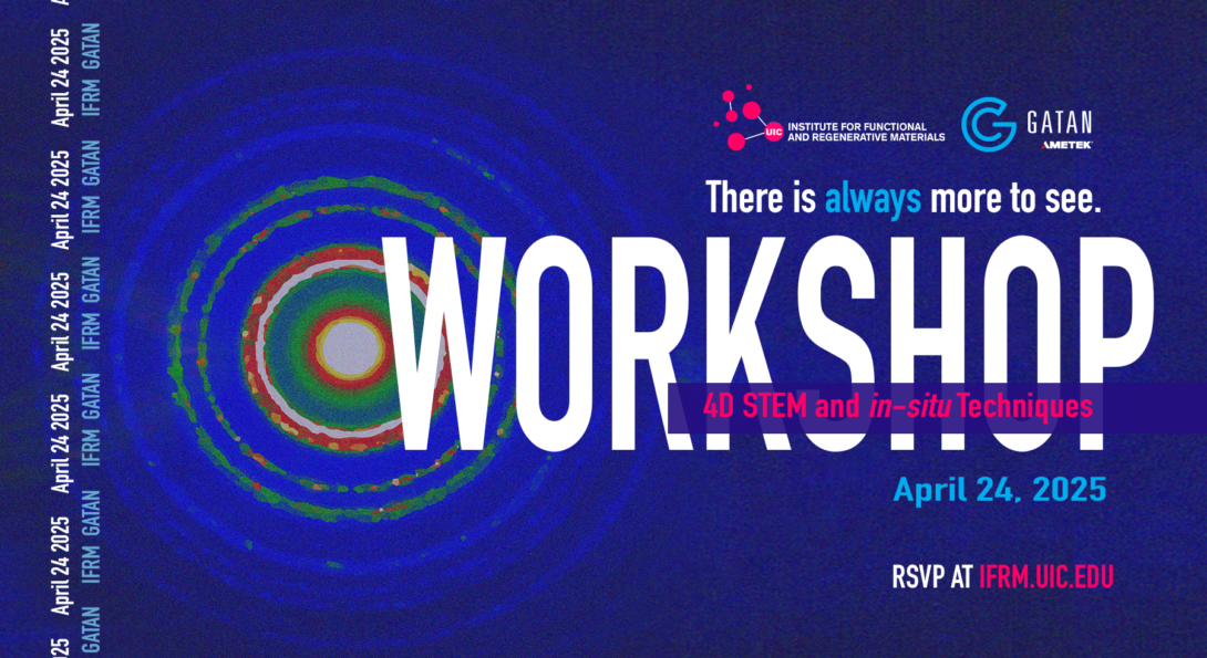
Join the UIC Institute for Functional and Regenerative Materials (IFRM) and Gatan for a 1-day workshop on in-situ and 4D STEM techniques with the Gatan ClearView camera. The event will feature TEM and STEM-focused talks from Chicago area microscopists and a demo of in-situ and 4D STEM data acquisition with the ClearView camera. The workshop will conclude with a seminar by David McComb (The Ohio State University).
Workshop Details
April 24, 2025 // 9:30 AM – 4:30 PM
UIC Science and Engineering South (Workshop Sessions)
UIC Lecture Center D5 (McComb Seminar)
How to Participate
Who Should Attend
The workshop is open to all interested in the field of functional imaging. This includes persons from industry, academia, and national laboratories. Students of all levels are also welcome to attend as attendees. Postdocs, graduate students, and advanced student researchers are also invited to submit posters to present during the workshop.
Workshop RSVP
RSVP is now closed. If you want to join remotely, please email Thomas at talaan@uic.edu for link.
Workshop Agenda
| 9:30 AM | Registration and Welcome |
| 10:00 AM | Gatan: Camera Technology and 4D STEM |
| 10:30 AM | Gatan Demo |
| 11:30 AM | Lunch |
| 12:30 PM | Arashdeep Thind (University of Illinois Chicago) |
| 1:00 PM | Danial Zanganeh (University of Illinois Chicago) |
| 1:30 PM | Chang Liu (University of Chicago) |
| 2:00 PM | Roberto dos Reis (Northwestern University) |
| 2:30 PM | Closing Remarks |
| 3:00 PM | Break |
| 3:30 PM | IFRM Seminar Series: David McComb (The Ohio State University) |
Invited Speakers and Abstracts
More bios coming soon.
Probing structure of functional oxides using 4D-STEM
Abstract
Four-dimensional scanning transmission electron microscopy (4D-STEM) provides a unique capability of simultaneously probing materials in real space and reciprocal space. 4D-STEM can accurately reveal epitaxial relationships, space-group symmetries, strain accommodation, and defects in a sample over 100’s of nanometers by sampling a much larger volume than typical atomic-resolution imaging. In this talk, I will show the role of epitaxial strain as a driver of ferroic transition in SrHfO 3 thin films grown by hybrid molecular epitaxy. Using 4D-STEM and other imaging and spectroscopy techniques, I will reveal the impact of high compressive strain in SrHfO 3 on its phase stability, octahedral tilts, and structural/chemical heterogeneities. Moreover, I will also show the layer-by-layer evolution of defect formation in Ruddlesden-Popper and infinite-layer nickelates, which has implications for high-temperature superconductivity.
About Dr. Thind / Profile
University of Illinois Chicago
Dr. Arashdeep Thind is a Postdoctoral Research Associate in Prof. Robert Klie’s group at University of Illinois Chicago. Arashdeep completed his PhD in Materials Science and Engineering at Washington University in St. Louis. He specializes in various electron microscopy techniques and first-principles modeling to investigate a variety of functional materials. He has received student awards from the Materials Research Society and the Microscopy Society of America and a postdoctoral award from the Postdoctoral Association at the University of Illinois Chicago.
Danial Zangeneh Talk
Abstract
Understanding phase stability in multivalent oxide cathodes is essential for advancing next-generation battery technologies. Here, we use in situ transmission electron microscopy (TEM) to directly visualize beam-induced structural evolution in MgCr1.5Mn0.5O4 at atomic resolution. Real-time observations reveal a transformation from the spinel to rocksalt phase, driven by cation redistribution and local chemical changes under electron irradiation. These results offer new insights into the dynamic behavior of complex oxides and highlight the importance of structural robustness in the design of durable Mg-ion battery materials.
About Zangeneh
University of Illinois Chicago
Danial Zangeneh is a Ph.D. candidate in the Nanoscale Physics Group at the University of Illinois at Chicago (UIC), working under the supervision of Professor Robert Klie. His research focuses on advanced electron microscopy and spectroscopy techniques for investigating materials at the atomic scale. He applies a variety of electron microscopy methods to systems such as magnesium-ion batteries, quantum dots, and superconductors, with a particular interest in structure–property relationships, beam-induced transformations, and interfacial phenomena. His work combines high-resolution imaging, in situ TEM, and spectroscopic analysis to uncover fundamental mechanisms that govern the performance and stability of energy and quantum materials.
Revealing Etching Trajectories and Heterogeneity of Nanocrystals in Liquid-phase Transmission Electron Microscopy
Abstract
Nanocrystals are inherently less stable than bulk materials due to their high surface-to-volume ratio, making them susceptible to decomposition under external stimuli. Understanding the degradation mechanisms of nanocrystals, particularly the pathways of structural transformation during deformation, is key to developing nanocrystals and devices with enhanced stability and robust structures. Here, we combine the real-space liquid-phase transmission electron microscopy (TEM) imaging and reciprocal-space 4D-STEM to study the morphological and structural evolution of metal and semiconductor nanocrystals, as well as the heterogeneous liquid environment. Our observations suggest the structure and morphology of nanocrystals could be controlled via external etching, shedding light on defect control and strain engineering in semiconductor nanocrystals and quantum dots for optoelectronic properties and devices.
About Dr. Liu / Profile
University of Chicago
Dr. Chang Liu is a postdoctoral researcher at the University of Chicago in Prof. Paul Alivisatos group. He has been working on in situ characterization of colloidal self-assembly and atomic structural dynamics of materials in heterogeneous environment using advanced electron microscopy such as liquid-phase TEM and 4D-STEM. Currently, his focus is on using in situ techniques to understand and provide insight on the structural, electronic, and vibrational properties of nanocrystals.
Physics-Guided Machine Learning in 4D STEM: A Multimodal Integration for Comprehensive Material Analysis
Abstract
This presentation introduces a framework integrating physics-guided ML with 4D STEM and spectroscopic techniques for advanced material characterization. Our approach employs PINNs to incorporate physical laws directly into EM data analysis. By embedding electron scattering theory and elasticity equations into NNs, we achieve improved accuracy in strain mapping and defect characterization compared to conventional methods. The framework enables true multimodal analysis by synergistically combining 4D STEM with EELS and EDS. Case studies on semiconductor heterostructures demonstrate correlations between structure, chemistry, and properties that remain hidden in traditional analyses. This physics-guided approach advances nanoscale materials characterization, providing deeper insights into complex material systems.
About Dr. dos Reis / Profile
The Ohio State University
Dr. Roberto dos Reis is Research Assistant Professor and Lecturer at Northwestern University. He conducted the implementation of 4D-STEM techniques and direct electron detectors at NUANCE Center, developing protocols for high-throughput data collection and analysis. He specializes in advanced data analysis in electron microscopy, focusing on AI-driven approaches and multimodal data integration. With expertise spanning quantum algorithms, AI/ML, and materials characterization, Dr. dos Reis has established innovative research programs bridging materials science with practical applications through electron microscopy.
Determining molecular functionality using electron energy-loss spectroscopy in the scanning transmission electron microscope.
Abstract
The need to characterize chemistry, structure and bonding with high spatial resolution has driven the exciting developments in electron energy-loss spectroscopy (EELS) in the scanning transmission electron microscope (STEM) over the last two decades. This has been achieved with the development of high-performance electron monochromators, improved spectrometer electron optics and introduction of new detectors with a high detector quantum efficiency (DQE). This has culminated in achievement of an energy resolution of 4 meV (32 cm-1) or less on a few STEM-EELS instruments. This level of performance is opening new opportunities for the study of both electronic and vibrational excitations using high resolution EELS in the scanning transmission electron microscope (STEM). One of the most exciting, and challenging, research opportunities for high resolution STEM-EELS is to identify functional groups in organic/biological systems and determine their spatial distribution. In the first part of this seminar, I will discuss recent research results demonstrating the use of high-resolution STEM-EELS to identify functional groups in polymers and to use this to spatially map such groups at the dentin-enamel junction (DEJ) to gain insight into biomineralization mechanisms. In many “soft” materials the damage caused by the electron beam limits the data quality that can be achieved, even with the use of “smart” scanning algorithms and machine learning optimizations. The use of cryogenic sample cooling has been employed to dramatic success in the determination of structure of proteins and macromolecules by cryoEM. This requires cryo-transfer of flash frozen samples into the microscope, a capability that, until recently, was not available on STEM-EELS systems capable of ultimate energy resolution. I will report the development of a cryo-transfer system for the monochromated aberration corrected (MAC-) STEM at Oak Ridge national Laboratory (ORNL) and will show early results on the study of several macromolecular systems by vibrational spectroscopy using cryo-STEM-EELS.
About Dr. McComb / Profile
The Ohio State University
David McComb, PhD, is the Founding Director of the Center for Electron Microscopy and Analysis (CEMAS), an Ohio Research Scholar and Professor of Materials Science and Engineering at The Ohio State University. A chemistry graduate from the University of Glasgow, David completed his PhD in Physics at the University of Cambridge. David is an expert in the development and application of electron energy-loss spectroscopy (EELS) as a sub-nanometer scale probe for chemistry, structure, and bonding. He has developed and implemented approaches to studying inorganic, organic, and molecular systems using electron microscopy techniques. He has extensive experience in the application of EELS to the study of problems in sold-state chemistry and materials science, including structural and compositional variations in high-k oxides, short range magnetic order in transition metal oxides, interfaces in fuel cells, photovoltaics, multiferroics and biomaterials. In recent years, he has focused his attention on developing and implementing approaches to studying organic and molecular systems using these methods. He has demonstrated that these methods can be used to obtain unique insights into materials such on polymers for organic photovoltaics, biomineralized tissues, amyloid plaques, wear particles in macrophage cells associated with metal hip implants, molecular fragments associated with degradation of carbon nanotubes in cells and mechanisms for early stage mineralization processes. He is a fellow of the Microscopy Society of America and the Royal Society of Chemistry and the Institute of Materials. In October 2011, he joined The Ohio State University as the founding director of the Center for Electron Microscopy and Analysis (CEMAS). This multidisciplinary facility drives the application of state-of-the-art electron microscopy techniques to strategic research challenges in the physical, engineering, life and medical sciences at Ohio State and beyond.